The geometry
PWHGMs combine a substantial hollow interior, an interconnected porous glass wall, and an accessible outer surface. These separate physical regions offer different opportunities to design pharmaceutical formulations.
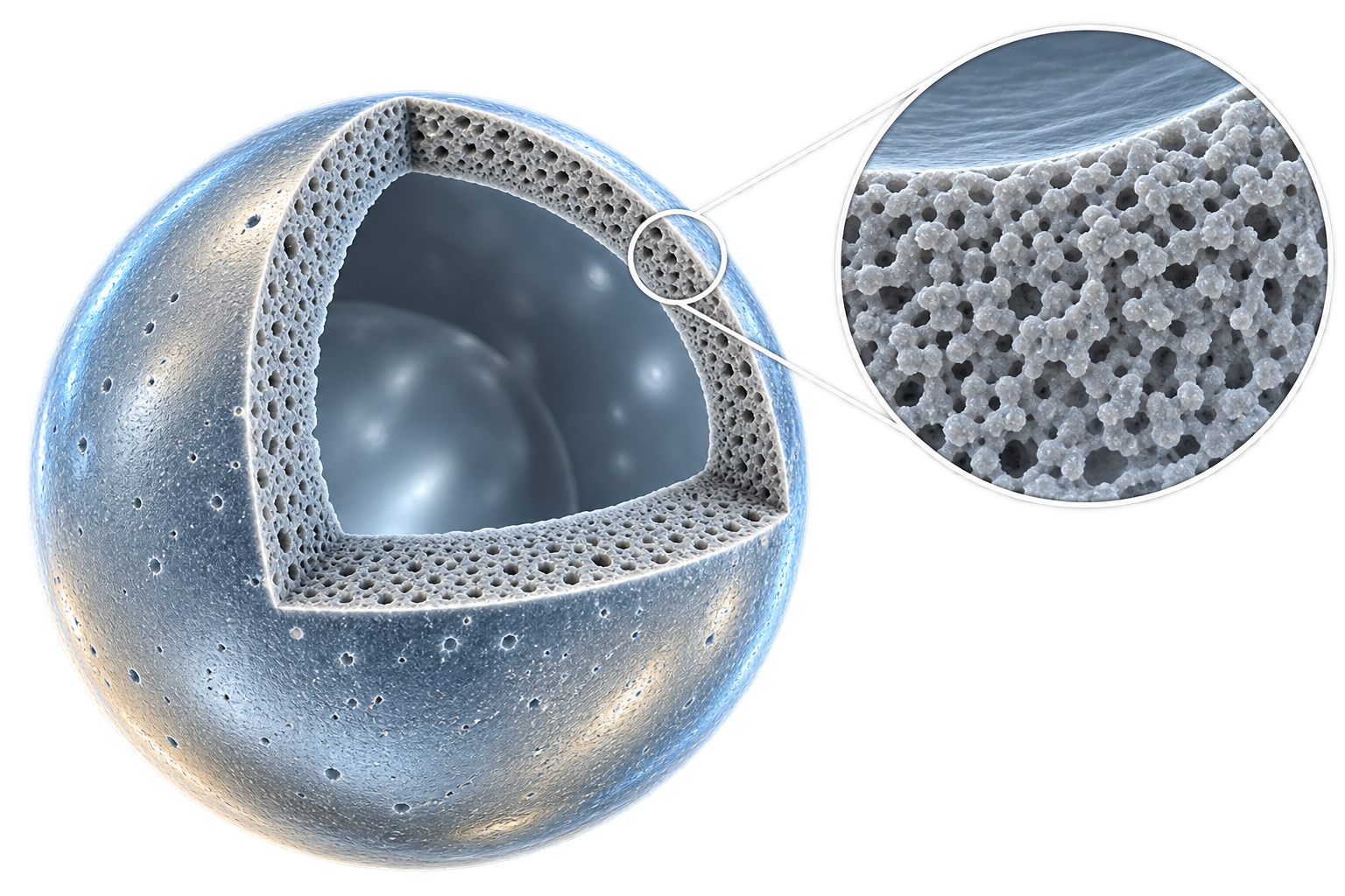
Conceptual cutaway showing the hollow interior and porous shell; not to scale.
What the particles actually look like
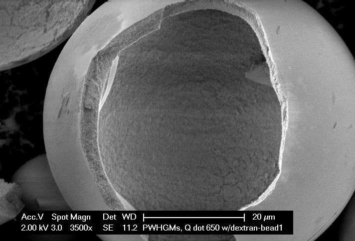
Intact microspheres imaged by scanning electron microscopy. The cutaway illustration above is a conceptual aid; the image below is genuine experimental imaging.
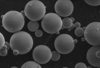
Think of the particle as three connected but distinct domains: the internal reservoir, the porous transport wall, and the external interface. This architecture distinguishes PWHGMs from silica systems that rely mainly on pore volume for cargo storage.
Reservoir versus porous matrix
In a conventional pore-based silica carrier, much of the available loading space lies within its porous network. In PWHGMs, a substantial hollow cavity provides a distinct storage region while the wall provides transport pathways. That distinction creates additional formulation design possibilities; it does not, by itself, establish superior loading or clinical performance.
Evidence boundary: Hollow architecture and nanoscale porosity are established structural features. Superior pharmaceutical loading, GI protection, and oral bioavailability require cargo-specific, comparative testing.
Want an accessible introduction? Listen to SpheroFill Insights: Tiny Bubbles, Big Possibilities ↗
From the shell to the nanoscale
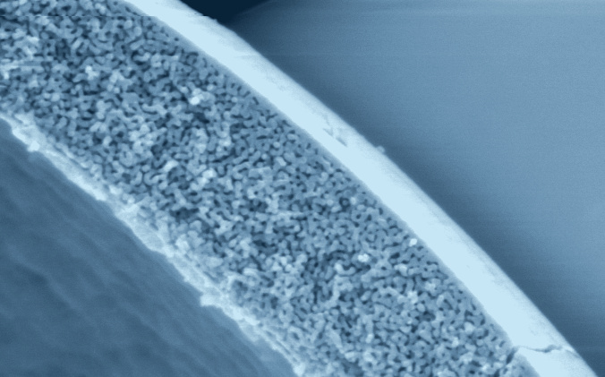
The ARC Hitachi micrographs below show cross-sectional wall structure, the hollow interior, and high-magnification porosity. The original instrument scale bars are preserved.
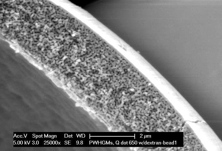
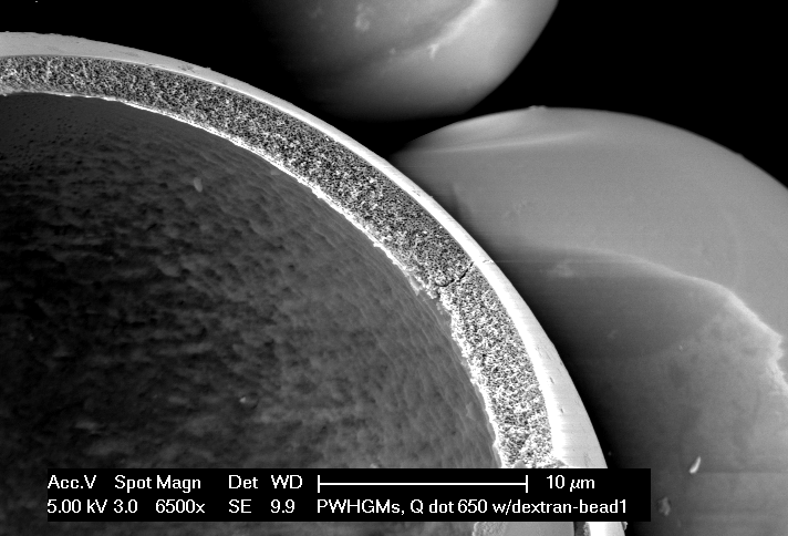
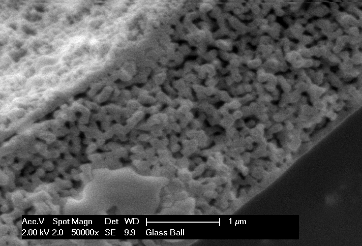
Measured architecture, not just an illustration.
Third-party analysis and SpheroFill characterization together show the platform's defining structural features. The values below come from specific historical characterization studies and illustrate the architecture that underlies the platform.
SRNL materials characterization
Third-party SRNL analysis documented wall micrographs, nitrogen adsorption and BET surface analysis, composition analysis, and particle-size distribution. One characterized historical sample measured 31.6 m²/g by BET.
SRNL-L3100-2012-00174 · Figures 6–8; appendices A–BPharmaceutical program characterization
NSF Phase I characterization found a mean diameter of 31.07 µm, a mean Feret ratio of 0.94 ± 0.04, a surface area of 16.5 m²/g, and an average pore width of 22.236 nm for the sampled material.
NSF SBIR/STTR Phase I final summary and characterization report · SpheroFill